The human brain is perhaps the most complex organic structure there is, says S.Ananthanarayanan.
The human brain has a hundred billion neurons, or nerve cells, and a trillion supporting cells. The brains cells train themselves to carry out tasks of cognition, reasoning, automation and memory, in ways that baffle computer scientists. But, complexity of its function apart, the living brain is a delicate organ, and study of its physical structure is not within easy reach.
Hsih-Yin Tan, Hansang Cho and Luke P. Le, from the National University of Singapore, the University of North Carolina at Charlotte, Sunkyungkwan University, South Korea, University of California at Berkley, Harvard Medical School and Brigham Women’s Hospital, Boston, in a Review Article in the journal, Nature Biomedical Engineering, recapitulate current laboratory methods to get a glimpse of the brain’s working. With the help of a scaffold that holds human nerve cells, these methods seek to create a model of a bit of brain, that allows conditions that lead to different brain diseases to be simulated and studied, the paper says.
The brain, being, quite literally, the nerve centre of the body, needs both protection and nourishment. In respect of nourishment, the brain receives blood supply - three quarters of a litre, which is 15% of the blood circulating in the body, every minute. The supply comes through two main sets of arteries, the carotid arteries to the front of the brain and the vertebral arteries that supply the rear parts of the brain. While the arteries are ‘autoregulated’, to keep up the supply of blood, the two artery systems are interconnected, so that supply continues even if one stream is blocked.
As for protection, the brain is kept separated, quarantined, one may say, from physical contact, even contact with the bloodstream. A system of tissue, known as the blood brain barrier, only permits what the brain needs, which is oxygen, glucose and nutrients, like amino acids, to pass from the blood to the brain, and not any infection, things dissolved in the blood, or some large molecules. Antibodies and immune cells are kept out, even therapeutic agents are not admitted. Within the brain, however, there is a system to receive, by diffusion, what the brain cells need, and to evacuate waste.
The paper explains that this system, which separates brain tissue from the bloodstream, while keeping steady the environment within the brain, is a key component of brain function. Damage to the system, mechanical or biological, results in the entry of harmful substances and disturbing of the internal balance, leading to damage and loss of brain cells. Conditions like formation of plaque in the blood vessels in the brain could increase the pressure and lead to rupture of blood vessels, which could also come about if there is physical injury, like a blow to the head.
These, and other brain conditions that arise from genetic or pathological causes, need invasive study to understand and analyse. As it is not practical to carry out such studies in a living brain, the recourse has been to animal studies. Animal studies are really not effective, the paper says. For instance, studies on the brains of mice have shown that that a gene that promotes a particular protein leads to conditions like Alzheimer’s disease, in mice. However, as the ratios of neurons and other cells in animals are different from the ratio in humans, and there are different immunological responses, the results of the gene and Alzheimer’s disease in mice cannot be carried over to draw conclusions about the human brain, the paper says.
There is hence a need, the paper says, for a functional model of the brain, which can more realistically represent the characteristics of the human brain and capture the way the brain responds to different kinds of stress. It is with such a ‘working model,’ the paper says, that we can understand brain mechanisms and the reasons for many brain diseases. This would bring brain research on par with other fields, like analysis of civil structures, or aircraft design, where parameters are varied and measured on models, as a means of discovering the behaviour of the real objects.
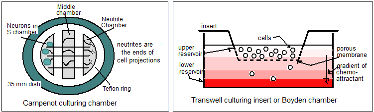
The paper carries out a review of the methods developed so far, to build a bit of the brain, using brain cell cultures, coaxed to form into desired structures with the help of mechanical scaffolds. Building on early methods of growing neuron cells on Petri dishes, were the Campenot Chambers, of specially shaped Petri dishes to guide the cell growth. An improvement was the Transwell culturing insert or the Boyden chamber, which separated cells by a porous membrane, the two sides to represent the ‘blood side’ and the ‘brain side’, which helped assess migration of cells across thin barriers.
More effective simulation of the 3-D structure of brain cells has been by growing cells in a 3-D microenvironment of a gel – to act as the extracellular matrix for cell growth. These structures have enabled recording spontaneous activity of neural and other cells and key mechanisms and pathways. However, there are limitations, the paper says, in simulating how brain cells relate to surrounding tissue – and these are better modelled in the brain organoid culture.
Organoids form when cells, derived from stem cells, organise themselves into 3-D structures. These cultures can be shaped to replicate a great part of the complexity of an organ, or specific aspects, such as the kind of cells that grow in the culture. “A simplified version of an organ produced in vitro in three dimensions that shows realistic micro-anatomy,” is one way it has been described. The advances in biomaterials engineering and genome editing techniques, to enable neural cells to be grown, have enabled such mini-brain models to be formed, the paper says. The techniques have become sophisticated and we are able to create personalised brain models, using the patient’s own body cells and re-enacting the path of disease progression.
Progress in organoids, with advances in 3-D printing techniques to build matrices to guide cell growth, has promoted brain modelling as a prime candidate for study of major neurological diseases. The development of Amyloid plaque, implicated in Alzheimer’s disease, is one. Parkinson’s disease, the second most common neurodegenerative disease, is another. And then, there is brain cancer. And next, what happens when there is traumatic injury to the brain. Mini brain modelling has the potential to develop precision and personalised therapy for diseases of the brain or the nervous system, the paper says.
------------------------------------------------------------------------------------------ Do respond to : response@simplescience.in------------------------------------------- 