Voice can act as the tissue probe, says S.Ananthanarayanan.
A technique used to image earth faults deep underground shows a way to peer into tissue of the human body.
The technique, which is described by Steve Beuve, Samuel Callé, Elise Khoury, Emmanuel Gilles Simon and Jean-Pierre Remenieras, from Université de Tours, Univerisité Bourgogne and the university hospital, Bourgogne, in France, in the journal, Applied Physics Letters, relates to the tissue of the thyroid gland. The thyroid gland can be affected by cancer. The current methods of detection of the cancer, however, do not locate the cancer precisely, and the detection of cancer can entail more discomfort than its treatment and cure. The new method, where the patient participates, leads to easier and more accurate detection.
The most common form of cancer of the thyroid gland, especially when detected early, is among the most treatable cancers. Detection is by physically feeling the area of the throat for the harder nodules that form, and with the help of ultrasound or x rays. If a tumour is found, this has to be followed by biopsy, or excision of a bit of the tumour, for lab tests, to see if the cells are malignant.
The excision is done by inserting a fine needle into the nodule and syringing out a sample. The procedure, known as fine needle aspiration, has often to be repeated, as it is only in some 5% of the trials that the cancer is detected.
A reason for the poor results could be that the ultrasound, or even sophisticated x ray methods, do not provide any supplementary information about the tissue in the places where they detect tumours. Ultrasound, for instance, does not create the contrast needed to make out live and dead tissue. The CAT scan, which uses x rays to image an organ in several ‘slices’ and puts together a 3D picture, is sensitive, but does not help make out malignancy.
The method now used to probe what is called soft tissue in the body, in different organs, is shear-wave elastography. Mechanical waves, like sound waves, that pass-through media, are compression waves, or longitudinal waves, where the to-and-from motion of material is along the direction of the wave. In the ripples on the surface of a pond, or a violin string that is plucked, in contrast, the motion is at right angles to the direction of the wave. These are called transverse wavess. Transverse waves can also arise when the surfaces of bulk material are exposed to vibration. Here, planes within the material move relative to each other, and these are called shear waves
The movement of longitudinal and transverse waves through the body of the earth, and over its surface, is routinely used to make out seismic fault lines, presence of water, or minerals or ores, etc., within the depths of the earth. In the case of tremors set off by movements in the earth’s crust, undersea volcanic action, etc, waves which travel, directly and after reflection at discontinuities within the earth, get detected at observation centres, to locate and assess the severity of the tremor. Waves could also be generated on purpose, usually by explosives, and the timing of detection at different observation points leads to information about the nature of the material at different depths under the earth’s surface.
The use of elastography to probe body tissue is an extension of the same principle. As the movement of shear waves through the body of tissue brings about compression and bending of the material, a shear wave would scatter waves of ultrasound that are passed though the tissue at the same time. If there are spots where the material is of greater stiffness, the shear waves would move faster when they pass through these portions, and this would show up in the ultrasound scattering pattern. The method is then a sensitive way of discovering non-uniformities, and hence possible malignancy, in soft tissue.
A development of the technique is not to use an external source of vibration of the surface of the tissue, to create the shear wave, but to rely on the body’s own physiological distortions, like the beating of the heart, expansion and contraction of blood vessels or respiration. The method is both non-invasive and sensitive, and by probing the tissue under study by ultrasound from different directions, an accurate map of the tissue distribution can be obtained. As there is no active source of vibration, the method is called Passive Elastography. The paper in Applied Physics Letters says that this technique has been applied to look for tumours in the thyroid, and the main arteries in the neck region are most suitable as sources of shear waves.
,/i>A factor that determines how effective this method can be is how strong the source of vibration is, in comparison with incidental disturbances, what is called the signal to noise ratio. In passive elastography, as the source of vibration is feeble, this could be a limitation. And then, the source of vibration is not what the investigator can control, and the investigation has to make the best of available sources.
The work reported in Applied Physics Letters moves forward, half-way in principle, but a lot in results, by harnessing the patient’s own voice to work as the source of vibrations. The carotid artery, which is right next to the thyroid gland, the paper says, vibrates at the rate of less than ten beats a second. A higher frequency, which would create shear waves of shorter wavelength, would be more suitable. And a stronger source of waves would improve the signal to noise ratio too.
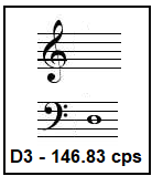
The paper reports that the vibrations of the vocal tract, when the patient vocalises a tone with a frequency of 150 cycles a second, produces robust shear waves that allow imaging of tissue with fine resolution. A tone at 150 cycles per second corresponds to something like D3, just to the left of the centre of the piano keyboard, an easy tone for a man or a woman to produce. And with this tone, using this method, which the authors call Vocal-Passive Elastography, the authors could reach a resolution of 150 microns, or about a sixth of a millimetre. We can see that shear waves at 150 cps would be more than 15 times shorter than the waves at 0-10 cps, which come from sources like heartbeat or the carotid artery.
An innovative discovery of a higher frequency vibrator, right at the spot, at the thyroid gland, for accurate identification of where to aspirate. Reducing the discomfort of the patient, and involving her in the procedure, with a singing lesson, of holding a note, to boot!
------------------------------------------------------------------------------------------ Do respond to : response@simplescience.in------------------------------------------- 